
Fig. 1.
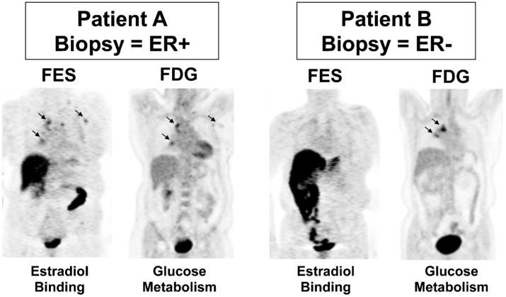
Imaging examples from two patients who underwent both FES and FDG scans prior to therapy. Left panel: Patient A (15-002) had mediastinal lesions appreciated by both FES and FDG. Right panel: Patient B (3-001) also had mediastinal disease clearly seen by FDG-PET, not visible on FES-PET. The core biopsy of a metastatic axillary lesion from Patient A showed ER+ breast cancer, while the needle biopsy of a vertebral lesion from Patient B showed ER- breast cancer.