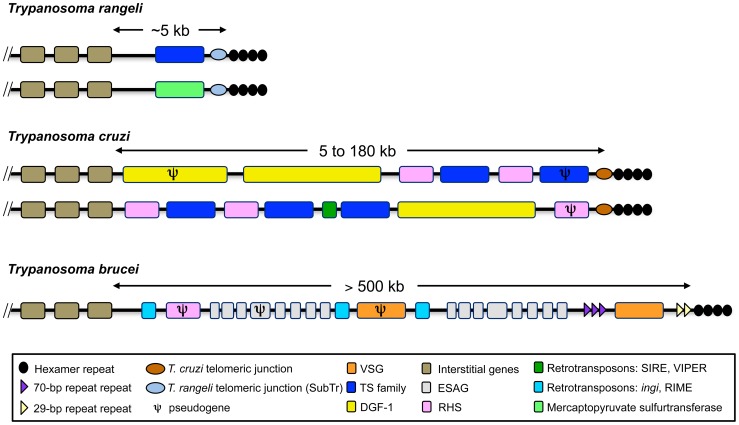
Figure 4. Representation of the telomeric and subtelomeric regions of Trypanosoma rangeli, T. cruzi and T. brucei.
The two types of telomeres identified in T. rangeli and two others representing the heterogeneity of T. cruzi chromosome ends are shown. The size of the subtelomeric region, which extends between the telomeric hexamer repeats and the first internal core genes of the trypanosomes, is indicated below each map. Boxes indicate genes and/or gene arrays. The maps are not to scale. The T. brucei and T. cruzi maps were adapted from [55], [98].

