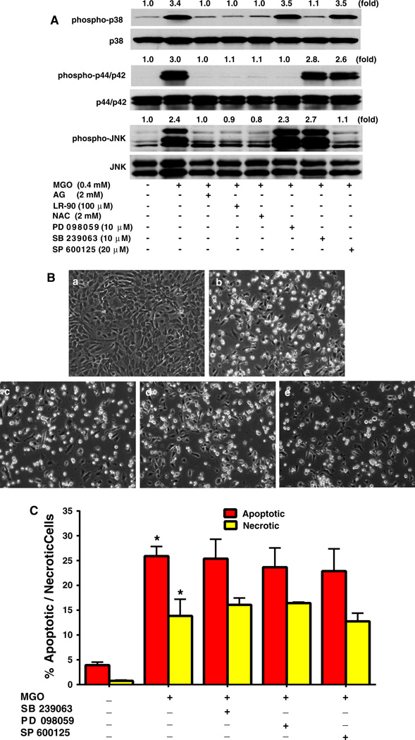
Fig. 6.
Effects of LR-90, AG and NAC on MAPK signaling pathways in HUVECs a Effects of LR-90 on MAPK signaling pathways. Representative Western blots of total and phosphorylated forms of MAPKs. Cells were pre-treated with test compounds for 1 h, and then stimulated with 0.4 mM MGO for 1 h. Numbers above each blot represent fold increase in protein expression of phosphorylated MAPKS relative to the control as quantified by densitometry and calculated with reference to the total forms of each MAPK. b Representative phase contrast photomicrographs of MGO-treated HUVECs with MAPK inhibitors. Cells were pretreated with test compounds for 1 h then co-incubated with a vehicle control; b MGO (0.4 mM); c MGO + 10 µM SB 239063; d MGO + 10 µM PD 98059; e MGO + 20 µM SP 600125, for 24 h. c Mean percentage of cells in early apoptosis or late apoptosis/necrosis as analyzed by flow cytometry. Data on graph were from three independent experiments (n = 6) and were analyzed by ANOVA followed by Bonferroni’s post hoc test (*p < 0.05 vs. vehicle control)

