
Fig. 2.
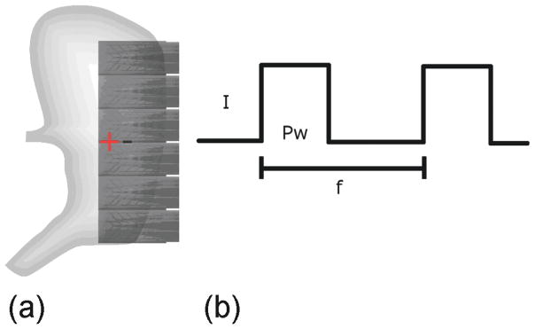
(a) Placement of the PCB electrodes. The “+” and “−” represent the location of the positive and negative leads of the stimulator, respectively. (b) Bipolar stimulus was used. The amplitude (I) for all stimulation trials was 2 mA and the pulse width (Pw) was 400 ms. This protocol was selected based on protocols that have previously been shown to successfully entrain gastric slow waves [20]. The frequency (f) of stimulation was 3.5 cpm (one pulse per 17 s).