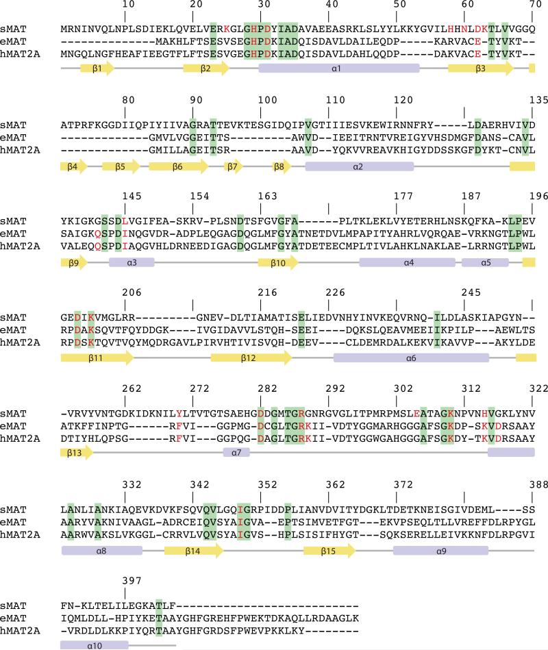
Fig. 2.
The structure-based sequence alignment of MAT from Sulfolobus solfataricus (sMAT), MAT from E.coli (eMAT) and human MAT (hMAT2A). Secondary structural features of sMAT are shown at the bottom. The numbering of the amino acids in the figure corresponds to sMAT. Identical residues between all three sequences are shown in green; identical residues between two sequences are shown in yellow. The critical residues involved in substrates binding are highlighted in red letters.

