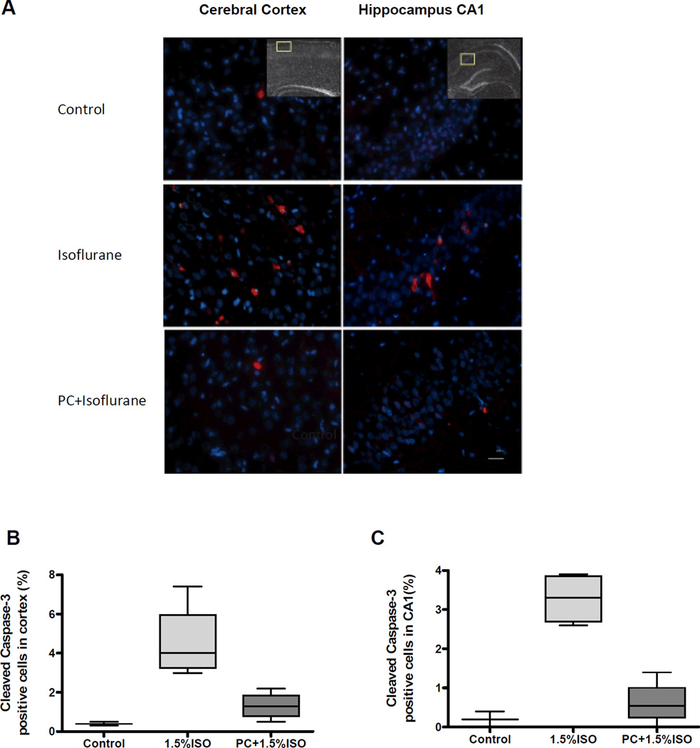
Figure 2. Isoflurane preconditioning significantly inhibits neuronal apoptosis induced by prolonged exposure to isoflurane in the cerebral cortex and hippocampus.
A) Representative images of immunostaining for cleaved caspase-3 in the postnatal day 7 (P7) rat cerebral cortex (left panels) and hippocampus (right panels) of controls (top panels), after isoflurane exposure (middle panels), and preconditioning (PC) with a brief anesthetic exposure before the isoflurane exposure (PC + Isoflurane) (bottom panels). Apoptotic cells are stained for caspase-3 (red) and total cells are stained with DAPI (blue). The insets indicate the areas sampled in the cortex and hippocampus for quantitation. Scale bar, 25 µm. B) Quantitative analysis of the percentage of cleaved caspase-3 positive cells in the cerebral cortex and C) the hippocampal CA 1 region. No significant differences were found in the number of caspase-3 positive cells in the cortex in the isoflurane (P=0.034) or PC + isoflurane (0.049) groups compared to controls or between the isoflurane and PC groups (P=0.021), or in the hippocampus (P=0.034, P=0.21, P=0.021 respectively). Data represent the mean of 3 adjacent brain sections per animal, n=4 animals for both experimental groups and n=3 for controls. Data are presented as box plots of the means with whiskers (min to max).

