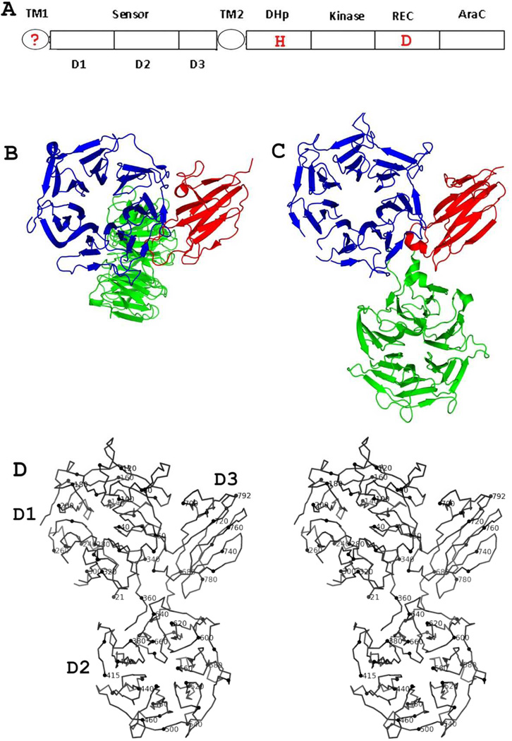
Figure 1. Structure of family 3 (HK3) histidine kinase receptors.
(A) Prototypical domain organization of HK3-type receptors. The question mark in TM1 indicates that it may not be present as a TM helix (see Supplemental Figure S1). Positions of signature two-component phosphorylation sites at histidine (H) and aspartic acid (D) residues are marked in red.
(B) Ribbon diagram of HK3 sensor domain BT4673S. The orientation views domain D1 into the top view [38] of the β-propeller domain. The coloring is done by domain: D1 (blue), D2 (green) and D3 (red).
(C) Ribbon diagram of HK3 sensor domain BT3049S. The orientation of the D1/D3 unit is as in Fig. 1B. The coloring is as in (B). Figs. 1C and 1D were generated using PyMol [69].
(D) Stereodiagram of the α-carbon backbone trace of HK3 BT3049S. Every 10th residue (modulo 10) is highlighted and every 20th residue is labeled.

