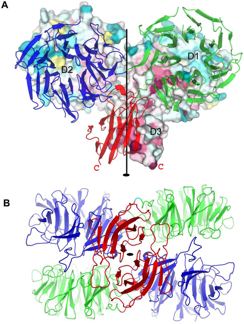
Figure 5. Dimeric structure of HK3 sensor protein BT3049S.
(A) View of the HK3 BT3049S dimer from within the plane of the membrane. The orientation is such that both β-propeller domains are viewed down their propeller axes. Propeller top sides [37] are in apposition at the reciprocal D1:D2′ interfaces. One protomer is shown as surface representation with residues colored based on its conservation through all HK3 homologs in B. thetaiotaomicron [73] and with domains D1, D2, and D3 labeled. The other protomer is shown as a ribbon diagram colored by domain: D1 (blue), D2 (green) and D3 (red). The C-terminus of each protomer is labeled as a red letter C. The quasi two-fold rotation axis is shown as a black line.
(B) View of the HK3 BT3049S dimer out from the membrane surface, from bottom of (A). The structure is shown as a ribbon diagram with coloring as for the ribbon protomer in C: D1 (blue), D2 (green) and D3 (red). The quasi two-fold rotation axis is shown as a black oval. The picture was made using PyMol [69].

