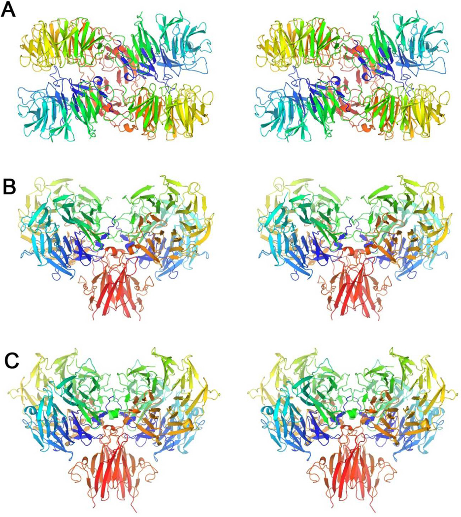
Figure 6. Stereoviews of productive B. thetaiotaomicron HK3 dimers.
(A) BT3049S dimer CD (PDBid 3V9F) viewed looking toward the membrane surface along the diad axis.
(B) BT3049S dimer CD viewed from the side, 90° from (A), with the putative membrane below.
(C) BT4663S dimer AB (PDBid 4A2L) viewed as in (B).
Each polypeptide chain is drawn in ribbon representation with spectral coloring, blue/green (N-D1) to yellow/orange (D2) to red (D3-C).

