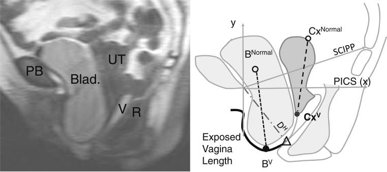
Fig. 2.
Mid-sagittal MRI at maximal straining and measurement scheme based with sacrococcygeal inferior pubic point (SCIPP) line-oriented axes; cervix location at maximal straining (CxV); most independent bladder point at maximal straining (BV); normal cervix location (CxNormal) and normal bladder rest location (BNormal) according to Summers et al. [11]. Δ transition point where the anterior vaginal wall (AVW) contacts the perineal body. DH, R rectum, UT uterus, V vagina, PB pubic bone, PICS Pelvic Inclination Correction System

