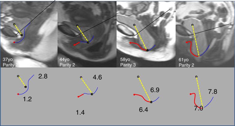
Fig. 3.
Examples of subjects’ MRIs and measurements. Open circle normal rest bladder location, filled circle subject bladder location at maximal straining with yellow dashed line representing the distance the bladder is below its normal rest location (number shown in cm). The red line indicates the portion of the vagina that is exposed (length shown in cm) and the blue line the portion of the vagina that is in contact with the posterior vaginal wall. The filled triangle represents the transition point where the anterior vagina loses contact with the posterior vaginal wall

