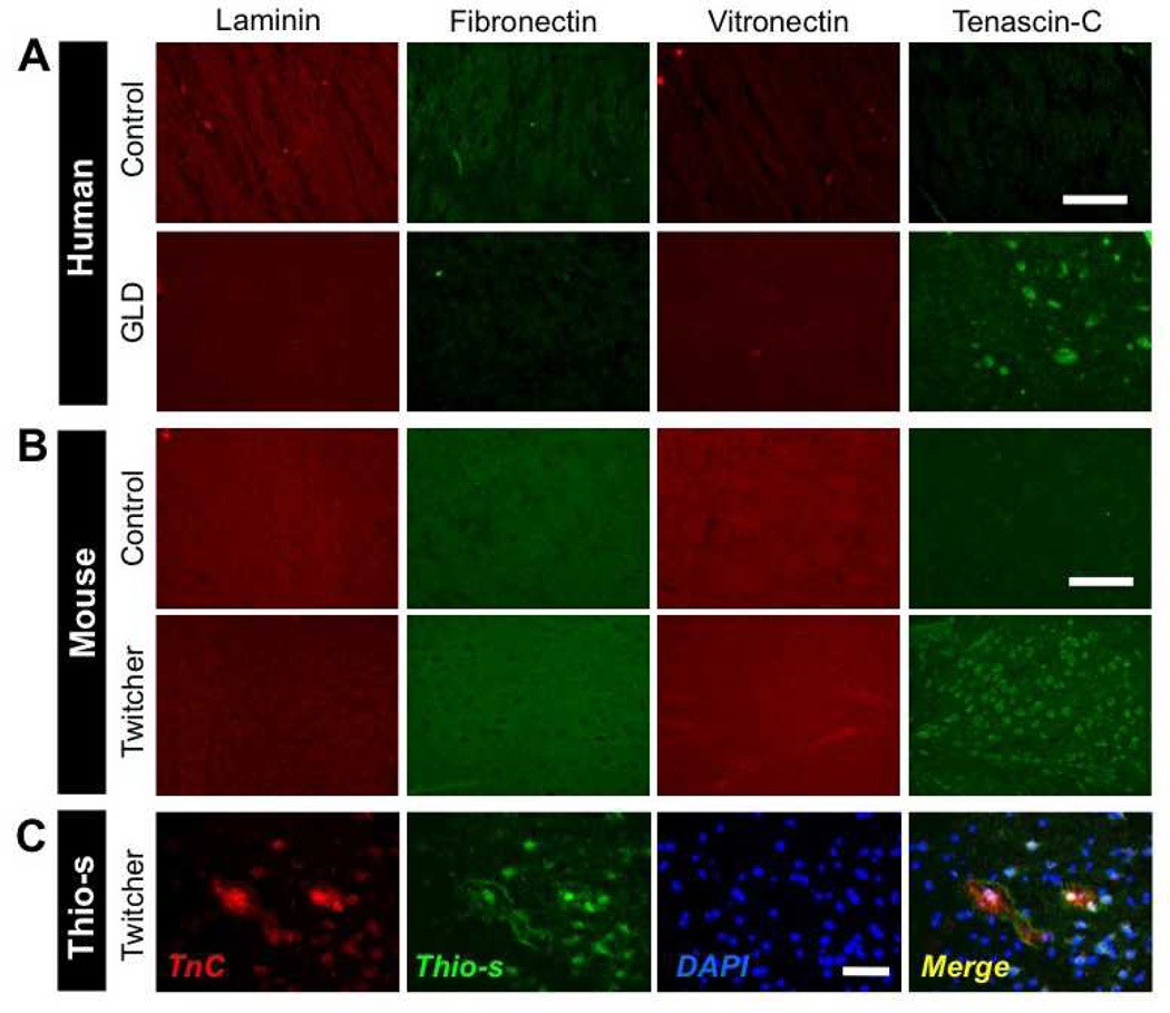
Figure 1.
Aberrant pattern of tenascin-C (TnC) expression in globoid cell leukodystrophy (GLD). (A) Immunohistochemical (IHC) analysis of the extracellular matrix (ECM) proteins laminin, fibronectin, vitronectin, and TnC in the midbrain of human infantile GLD patients (bottom row) and age-matched control human subjects (top row). (B) IHC analysis of ECM proteins in the midbrain of the P30 twitcher mouse model of GLD (bottom row), and age-matched littermate wild type control mice (top row). Note the intense punctate pattern of TnC immunoreactivity in human and murine GLD tissues vs. that in control subjects. (C) TnC immunoreactivity colocalizes with Thioflavin-S positive staining in the hippocampus of P30 twitcher mouse brain as visualized by IHC for TnC and Thioflavin-S staining. Nuclei are stained with DAPI (blue). Two to 4 specimens were analyzed. Scale bar: A, 250 µm; B, 150 µm; C, 100 µm.

