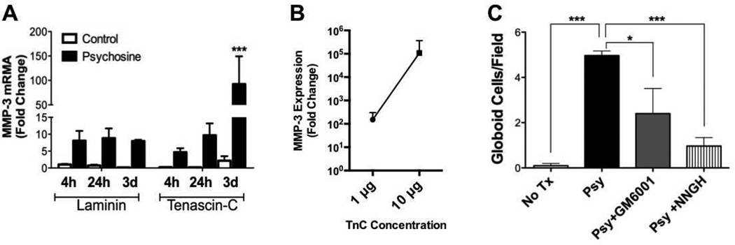
Figure 3.
Matrix metalloproteinase-3 (MMP-3) mediates globoid cell formation in response to psychosine. (A) Primary glial cell cultures, which consisted of astrocytes and microglia exclusively, grown on either laminin- or tenascin-C (TnC)-coated plates, were challenged with psychosine for 4 hours, 24 hours, or 3 days. Total RNA were isolated, reverse-transcribed into cDNA, and MMP-3 mRNA expression was analyzed by qRT-PCR. Data represent mean ± SE of fold change normalized to laminin-treated 4-hour samples. (B) Analysis of TnC concentration dependent effect on MMP-3 expression following 3 days of psychosine treatment. (C) Primary glial cultures grown on TnC-coated coverslips were treated with either psychosine (Psy; black bar), psychosine with the pan-MMP inhibitor, GM6001 (12.5 µM; gray bar) or the MMP-3 specific inhibitor, NNGH (0.1 µM; striped bar), for consecutive 7 days. Cells were fixed and immunostained for microglial marker, Iba-1 and DAPI for nuclei. Four to 6 visual fields under 20x magnification were randomly chosen and globoid cells were counted. Numbers of globoid cells were significantly decreased when GM6001 or NNGH was applied in culture in conjunction with psychosine treatment (Two-way ANOVA; interaction: **p < 0.0099; treatment: ***p < 0.0002). Data represent mean ± SE. N = 3/condition.

