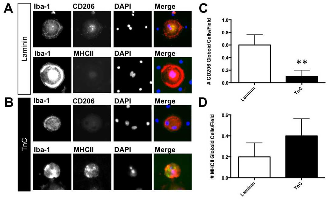
Figure 4.
Tenascin-C (TnC) changes M1/M2 phenotype in psychosine-treated microglia/globoid cells. (A, B) Isolated microglial cultures were grown on either laminin (Lm) (A) or TnC (B) and treated with psychosine for 7 days. Expression of M1 and M2 markers (major histocompatibility complex II [MHCII], CD206, respectively) was identified by immunocytochemistry. Representative images show that with microglia plated on Lm predominantly express CD206 but not MHC II (A), while cells grown on TnC predominantly express MHC II, but not CD206 (B). (C, D) Data represent percent of marker-positive cell staining of total cell number/field (n = 5–8 fields per treatment) (mean ± SE), **, p < 0.01. Scale bar: 20 µm.

