Figure 5.
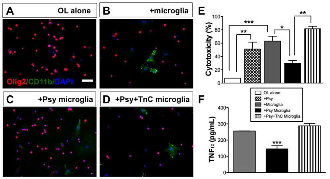
Globoid cells grown on tenascin-C (TnC) are toxic to oligodendrocytes. Isolated microglia treated with psychosine (+Psy), or grown on TnC and treated with psychosine (+Psy+TnC), or vehicle for 7 days were then co-cultured with oligodendrocytes for 4 subsequent days. Oligodendrocyte cultures were about 50% myelin basic protein (MBP)-positive at the time of co-culture. (A-D) Representative photomicrographs of immunocytochemistry for Olig2 (red) and CD11b (green) counterstained with DAPI (nuclei; blue) in cultures of oligodendrocytes (A), oligodendrocytes co-cultured with microglia (B), oligodendrocytes cultured with psychosine-treated microglia that were grown on a laminin substrate (C), or oligodendrocytes cultured with psychosine-treated microglia that were grown on a TnC substrate (D). Scale bar = 100 µm. (E) Analysis of cell death under each condition was measured by lactose dehydrogenase assay. Cytotoxicity is expressed as percent of full kill control (0.1% sterile Triton X-100). Psychosine-induced globoid cells without TnC (black bar) had minimal toxicity in co-culture to oligodendrocytes vs. untreated microglia (gray bar), whereas TnC-grown psychosine-treated microglia exhibited significant toxicity (striped bar). (F) Level of tumor necrosis factor (TNFα) released from isolated microglia treated with psychosine, TnC grown microglia treated with psychosine, or vehicle control (7 days) was measured by ELISA. Note that psychosine-treated microglia (black bar) exhibited the least amount of TNFα release vs. untreated or TnC-exposed psychosine-treated microglia (gray, striped bar, respectively). One-way ANOVA, p < 0.001; where, *p < 0.05; **p < 0.01; ***p < 0.001. N = 3/condition.

