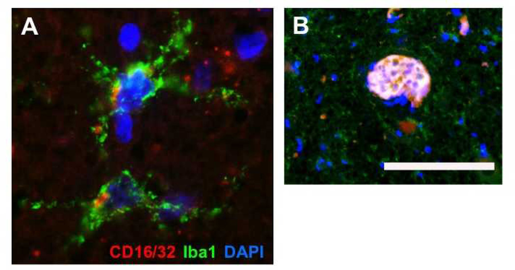
Figure 6.
Identification of M1 polarized microglia and Globoid Cells in globoid cell leukodystrophy (GLD). (A) Immunohistochemistry performed on brainstem tissues of an infantile GLD case identified multiple microglial cells expressing both Iba-1 and the M1 polarization marker CD16/32. (B) Within a white matter lesion with evidence of intense microgliosis, a multi-nucleated globoid cell was also identified that was immunopositive of both Iba-1 and CD16/32. Scale bar in B = 29 µm for panel A and 166 µm for panel B.

