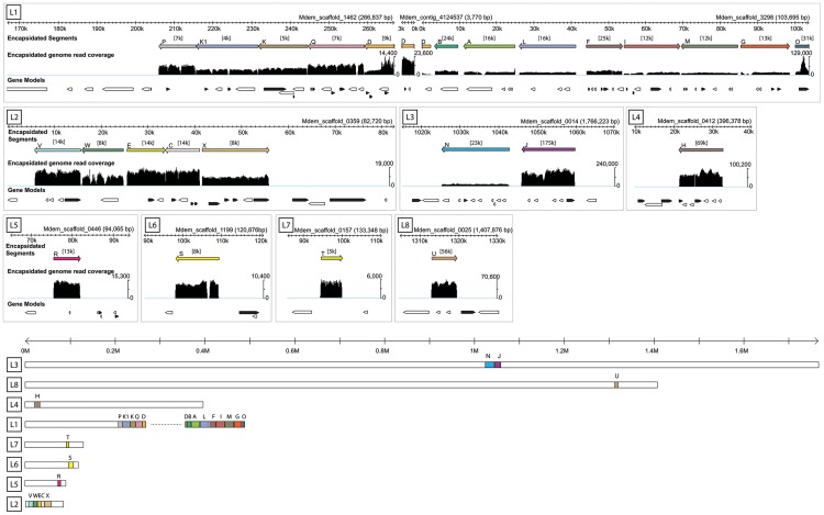
Figure 1. Genomic organization of MdBV proviral segment loci.
The upper part of the figure presents the eight proviral loci identified (L1–L8) and the corresponding M. demolitor genome scaffolds where they are located. Only the portion of the scaffold where the proviral locus resides is shown. For each locus, the upper scale bar in kilobases (k) names the scaffold(s). Below the scale bar, colored bars indicate the segments identified by deep sequencing DNA from MdBV virions (Encapsidated segments) and their corresponding orientation and location in a given locus as a proviral segment in the M. demolitor genome. Average coverage in thousands of reads is indicated above each segment in brackets, while below each segment is shown read coverage per nucleotide relative to the scale indicated to the right of the graph. Gaps in read coverage indicate regions flanking individual proviral segments that are not amplified, excised and packaged into virions. The gap seen in segment S (L6) is due to a region of N's in the reference sequence. Below each MdBV proviral segment is shown predicted genes in forward (black) and reverse (white) orientation. Individual introns, exons and untranslated regions are not shown. The lower part of the figure shows each scaffold containing MdBV proviral loci in their entirety. Scaffolds are drawn to scale and organized from largest (L3) to smallest (L5). Scale bar is in megabases (M).

