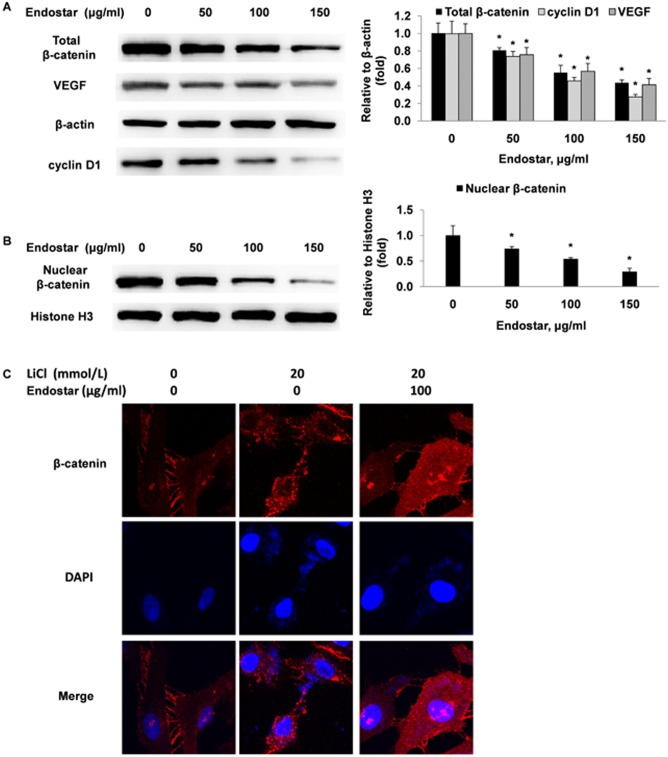
Figure 6. Endostar suppresses the expression of nuclear and total cellular β-catenin, cyclin D1 and VEGF in HUVECs.
(A and B) HUVECs were incubated with different concentrations of Endostar for 24 h. The whole-cell extracts and the nuclear extracts were prepared and analyzed by Western blotting and probed with specific antibodies. *P<0.05 versus the control group. (C) HUVECs were treated with or without 20 mmol/l LiCl (a dose known to activate β-catenin) in the presence or absence of 100 µg/ml Endostar. After 24 h, the expression and cellular localization of β-catenin (red) was determined by immunofluorescence analysis. The nucleus was stained with DAPI (blue).

