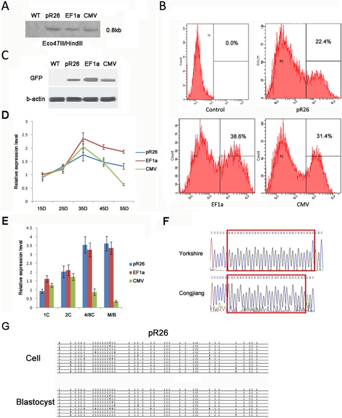
Figure 2. GFP expression driven by the pRosa26 promoter.
(A) Southern blot of the transgenic cell lines. Expected bands of 0.8 kb were detected after Eco47III/HindIII digestion. (B) Flow cytometry analysis of the transgenic cell lines. (C) GFP expression in the transgenic PFFs detected by Western blot. (D) GFP expression in transgenic PFFs over a long term culture up to 55D detected by Q-PCR. (E) GFP expression in transgenic cloned embryos detected by Q-PCR. (F) pR26 sequence of Congjiang minpig and Yorkshire pig. There is a lack of GGC in pR26 sequence of Congjiang minpig compared to Yorkshire pig. (G) DNA methylation status of pR26 in transgenic PFFs and cloned blastocysts. The methylation status was detected by the bisulfite sequencing. Methylated and non-methylated CpG dinucleotides of each clone are illustrated with closed and open circles, respectively.

