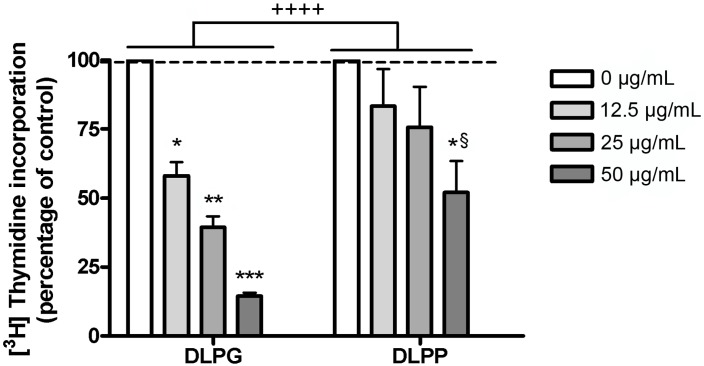
Figure 5. Comparison of the Effects of DLPG and DLPP on Keratinocyte Proliferation.
Near-confluent to confluent keratinocytes were treated for 24 hrs with the indicated concentrations of liposomes composed of DLPG or DLPP, prepared via bath sonication of the phopholipid in SFKM. [3H]Thymidine incorporation into DNA was then determined. Values represent the means ± SEM of more than 3 separate experiments performed in duplicate; **p<0.01, ***p<0.001 versus the control value (0 µg/mL); §p<0.05 versus the corresponding concentration of DLPG; ++++ p<0.0001 as indicated. [3H]Thymidine incorporation into DNA in the control was 50,400±7,500 cpm/well, and 53,300±6,400 cpm/well for DLPG and DLPP, respectively.

