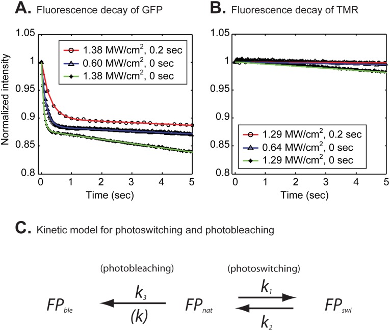
Figure 1. Fluorescent decay curves of H2B-GFP (A) and H2B-Halo-TMR (B) during time lapse imaging of entire nuclei in live cells.
The GFP tag exhibited a bi-phasic decay with three different combinations of the laser power and delay time between images (A). Colored curves indicate fits to the data with a bi-exponential decay model. According to the model (C), fluorescent molecules  can convert at a rate
  into a photoswitched dark state
  and then revert to the fluorescent state at a rate
  . Fluorescent molecules can also bleach irreversibly to a dark state
  at a rate
  . In contrast to the GFP tag, the TMR-Halo tag exhibited a monophasic decay that was well fit by a single exponential (colored curves) (B). Fitting parameters are shown in Table 1.

