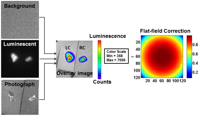
Figure 2. ROI selection on IVIS-200 images.
Three images–background, luminescent, and photograph were collected using IVIS-200 to create an overlay image. Then, a region of interest (ROI, shown with circle) was selected on the left carotid (LC) and right carotid (RC) arteries. The overlay image was corrected for field flatness, which was specific to the camera used in the IVIS-200 imaging system.

