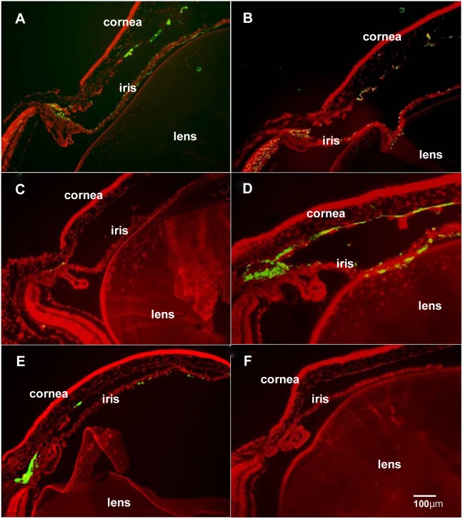
Figure 2. Intraocular localization of GFP delivered by intravitreous injections.
Ad5-GFP vectors transduced corneal endothelial cells, trabecular cells and iris cells (A). Ad5+GFP vectors transduced corneal endothelial cells, trabecular cells and iris cells (B). Ad35-GFP vectors transduced travecular cells alone (C). Ad35+GFP vectors transduced corneal endothelial cells, trabecular cells and iris cells (D). Ad28-GFP vectors transduced corneal endothelial cells, trabecular cells and iris cells (E). An Ad28-Null injected eye showed no GFP fluorescence in any ocular cells (F). Scale bar = 100 µm.

