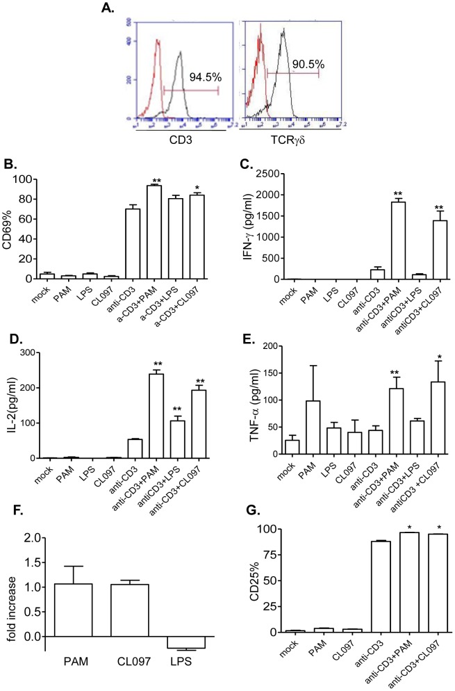
Figure 1. The effects of TLR2, 4 and 7 agonists on anti-CD3- treated murine γδ T cells.
A, Flow cytometry analysis of splenic γδ T cells stained with antibodies to TCRγδ and CD3. B–E, splenic γδ T cells were cultured with anti-CD3 with or without TLR agonists. Cells were harvested at 48 h post-stimulation and examined for CD69 expression (B), and IFN-γ (C), IL-2 (D) and TNF-α (E) production in culture supernatant. F. In vitro T cell proliferation assay. CFSE- labeled γδ+ T cells were cultured for 48 h in the presence of anti-CD3 with or without TLR agonists. Data shown are fold of increase of T cell proliferation compared to anti-CD3 treated cells. G. CD25 expression. Data are presented as means ± SEM, n = 4–7. ** P<0.01 or * P<0.05 compared to anti-CD3- treated cells. Data presented are one representative of at least four similar experiments.

