
Figure 1. Vacuolar change in ARQ/ARR sheep.
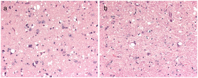
(A) Section of midbrain at the level of the superior colliculus from sheep 050 with multifocal neuronal vacuolation and extensive spongiform change in neuropil. (B) Section of brainstem at the level of the obex from sheep 055 with large intraneuronal vacuoles. Hematoxylin and Eosin Bar = 50 µm.