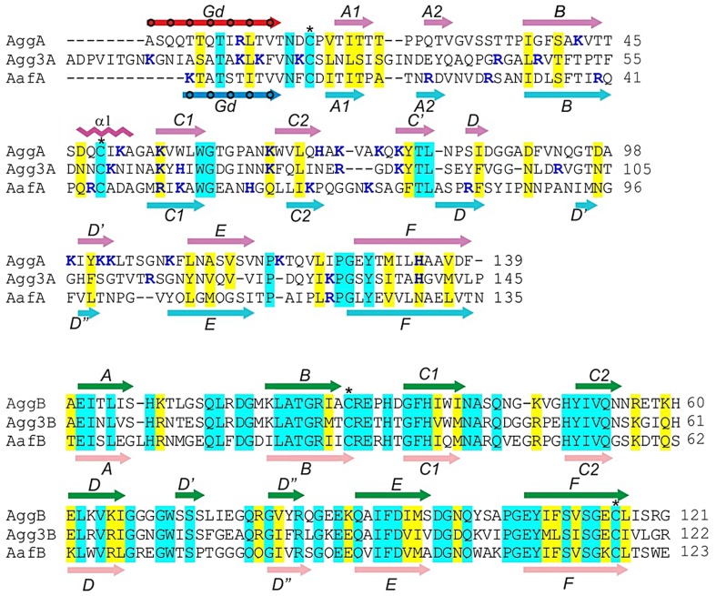
Figure 1. Sequence alignment of the major (AggA, AafA and Agg3A) and minor (AggB, AafB and Agg3B) subunits of aggregative adherence fimbriae (AAF) type I, II and III.
Secondary structure elements of AggA, AafA, AggB, and AafB core structures are shown in magenta, cyan, green, and pink respectively, whilst the donor strands in AggA and AafA (Gd) are shown in red and blue, respectively. Donor residues occupying pockets of the acceptor cleft are indicated with circles. Amino acid identities and similar residues are indicated by background shading in cyan and yellow, respectively. The donor residues, occupying pockets of the acceptor cleft are indicated with circles. Positively charged residues are shown in bold and painted in blue. Cysteine residues involved in disulfide bonds are indicated with stars. CLUSTALW alignment of sequences was modified based on superposition of structures of the donor strand complemented (DSC) subunits AggA and AafA and AggB and AafB (this study).

