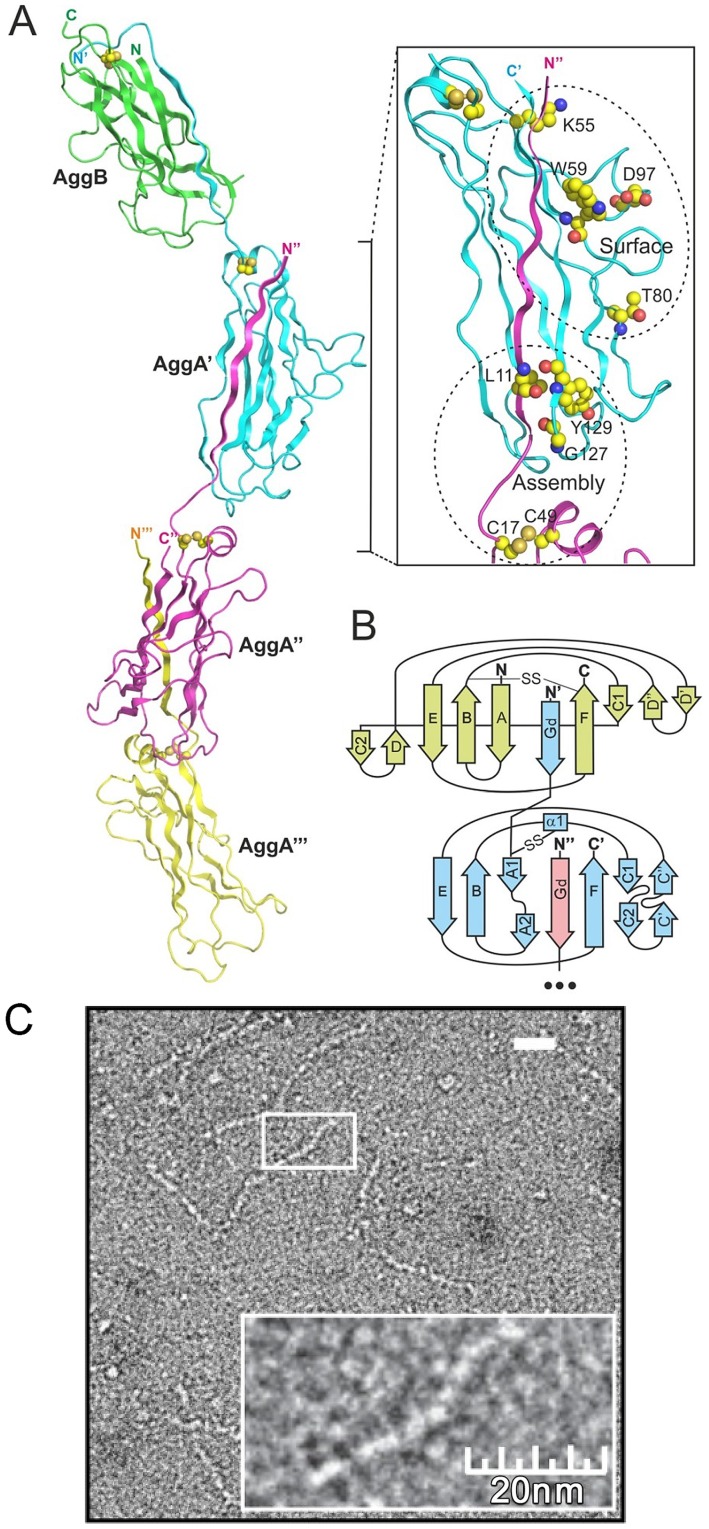
Figure 3. AAF architecture.
(A) Model of AAF/I constructed based on the crystal structures of DSC subunits, AggAdsA and AggBdsA, and the crystal structure of the F1 antigen mini-fiber [25] (cartoon diagram). The fiber contains a single copy of the AggB subunit (green) at the tip of a polymer of the AggA subunits (a fragment containing three AggA subunits is shown). The insert shows localization of conserved residues in the structure of the fiber. (B) Topology diagram of the AAF/I fiber. (C) Negative stain transmission electron micrographs of diluted AAF/II fimbriae isolated from enteroaggregative E. coli strain 042.

