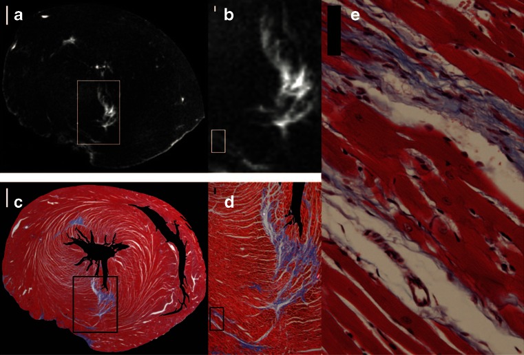
Fig. 2.
Histology and late gadolinium enhancement images in a rat model. Late gadolinium enhancement (LGE) tracks fibrosis to nearly the cellular level in this post infarction ex-vivo rat heart. LGE images were taken ex-vivo at 7 Tesla with a 3D gradient echo sequence (a and b) were compared with histological sections stained with Masson trichrome (c-e). Adapted from Schelbert et al. Circulation CVI 2010 [19••]

