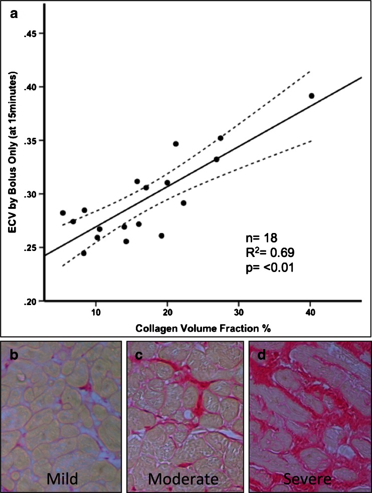
Fig. 3.
Histological validation of diffuse fibrosis in severe aortic stenosis. Top panel (a) showing correlation of extracellular volume (ECV) measured by CMR with histological collagen volume fraction in patients with severe aortic stenosis. The dashed lines represent 95 % confidence intervals. Bottom row (b-d) showing histology samples of mild (b), moderate (c) and severe (d) diffuse myocardial fibrosis. Picrosirus red stains collagen (fibrosis) red/pink, and counter stains myocytes yellow. Collagen volume fraction (%) was calculated by automated subtraction of these 2 color stains. Adapted from White et al. JACC CVI 2013 [47]

