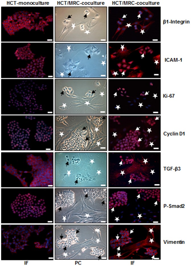
Figure 1. Intensive crosstalk between CRC and MRC-5 cells in the tumor microenvironment.
HCT116 cells (arrows) were either cultured alone or were co-cultured with MRC-5 cells (*) at a ratio of 1∶1 for three days on glass plates in monolayer and fixated with methanol. For immunolabeling cells were incubated with primary antibodies against β1-Integrin, ICAM-1, Ki-67, cyclin D1, TGF-β3, p-Smad2 and vimentin) followed by incubation with rhodamine-coupled secondary antibodies and counterstaining with DAPI to visualize cell nuclei. Images shown are representative of three different experiments. PC: phase contrast; IF: immunofluorescence; Magnification 400×; bar = 30 nm.

