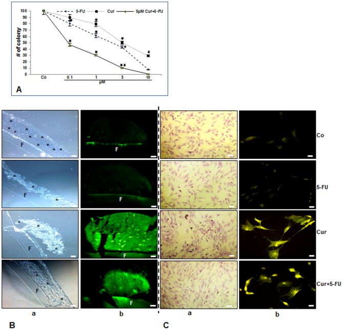
Figure 2. Toxicity of 5-FU, curcumin and the combination treatment on HCT116/MRC-5 cells and cellular uptake of curcumin in these cells in high density and monolayer tumor microenvironment co-culture.
A: Quantification of the number of colonosheres was achieved by counting the number of spheroid colonies from 10 microscopic fields in the high density microenvironment co-cultures. Cultures were either left untreated (Co) or were treated with 5-FU (0.1, 1, 5 or 10µM), curcumin (0.1, 1, 5 or 10µM) or were pretreated with curcumin (5µM) for 4 h, and then exposed to 5-FU (0.1, 1, 5 or 10µM) for 10 days and evaluated by light microscopy. Values were compared with the control and statistically significant values with p<0.05 were designated by an asterisk (*) and p<0.01 were designated by an asterisk (**). Toluidine blue staining profile (2B/C, a) and cellular curcumin uptake (2B/C, b) of HCT116 (B) in high density and in MRC-5 (C) in monolayer co-culture. Tumor microenvironment co-cultures were either left untreated (Co) or were treated with 5-FU (5µM) (5-FU), curcumin (5µM) (Cur) or were pretreated with curcumin (5µM) for 4 h, and then exposed to 5-FU (0.1µM) (Cur+5−FU) for 10 days and evaluated under a light or fluorescent microscope. Images shown are representative of three independent experiments. F = Filter. (*) = HCT116 colonosheres. Magnification 4B, 4Ca: 200x, 4Cb: 400x; bar = 30 nm.

