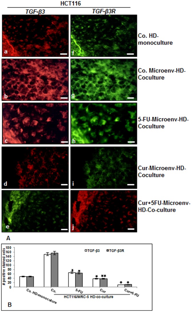
Figure 6. Curcumin or 5-FU suppresses TGF-β3 and TGF-βR expression in CRC cells in high density tumor microenvironment co-culture.
A: High density mono-cultures of HCT116 cells were left untreated, high density tumor microenvironment co-cultures of HCT116/MRC-5 cells were either left untreated, or treated with 5-FU (5µM), or with curcumin (5µM) or pre-treated with curcumin (5µM) for 4 h, and then exposed to 5-FU (0.1µM) for 10 days. The cultures were subjected to immunofluorescence labeling with primary antibodies for TGF-β3 (a-e) and TGF-β3R (f-j) followed by incubation with rhodamine- or FITC-coupled secondary antibodies. Images shown are representative of three different experiments. Magnification 400×. bar 30 nm. B: To quantify the amount of TGF-β3 and TGF-βR-positive cells in high density cultures described above, 200 cells from 15 microscopic fields within the stained slides were counted. The results are provided as the mean values with S.D. from three independent experiments. Values were compared with the control and statistically significant values with p<0.05 were designated by an asterisk (*) and p<0.01 were designated by an asterisk (**).

