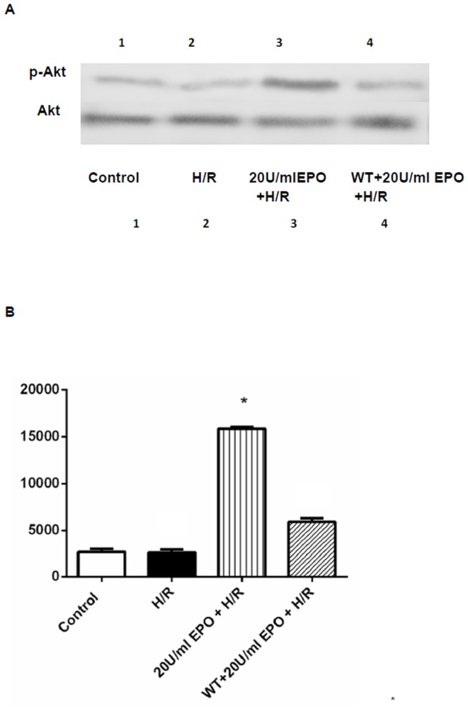
Figure 8. Western blot analysis demonstrating the effect of EPO on AKT.
The effect of EPO on p-Akt was determined using Western blot. H9C2 cells were isubjected to H/R with or without pre-treatment with 20 U/ml EPO for 24 hrs and incubated with 1 µM of Wortmannin 30 mins before EPO treatment. (A) Samples treated with EPO showed increase in phosphorylation of Akt (p-Akt). Akt remain unaltered and demonstrate equal protein loading in all lanes. (B) Represent the quantization of western blot, which indicates the increase in p-Akt levels Data are presented as means ± SEM of the ratios from three independent experiments * denotes p<0.001 for analyses compared to H/R.

