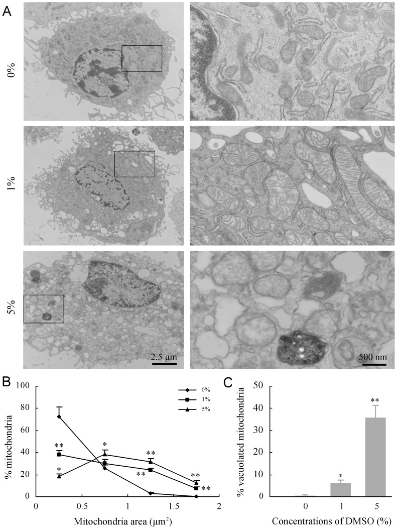
Figure 2. Effects of DMSO on substructures of cultured mouse cortical astrocytes.
(A) Representative transmission electron micrographs showing substructural morphology of astrocytes after treatment with different concentrations of DMSO for 24 h. The disruption of mitochondria integrity becomes more severe with increased DMSO concentrations. Fragmentation of the nucleus, with condensation and margination of nuclear chromatin, was frequently observed in astrocytes exposed to 5% DMSO. (B) Quantitation of mitochondrial cross-sectional area. The results confirmed DMSO-induced mitochondrial swelling, with a significant rightward shift in the mitochondrial area cumulative frequency curve, relative to untreated control. (C) The quantitative analysis showed increases in the percentage of mitochondrial vacuolization in astrocytes treated with DMSO in a dose-dependent manner. Data are shown as a mean ± SEM of five independent experiments. *P<0.05 and **P<0.01 versus control group.

