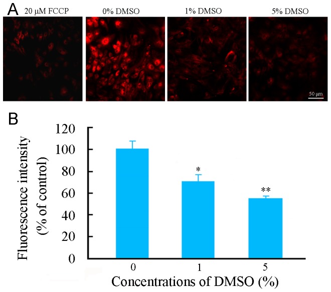
Figure 3. Effects of DMSO on ΔΨm of cultured mouse cortical astrocytes.

(A) Representative micrographs showing ΔΨm, revealed by TMRE fluorescent staining, in astrocytes treated with various concentrations of DMSO for 24 h. The left first micrograph is a positive control for depolarized mitochondria by incubated with 20 µM FCCP, an uncoupler of electron transport and oxidative phosphorylation, for 10 minutes prior to staining with TMRE. (B) The quantitative analysis revealed that TMRE fluorescence intensity was decreased in astrocytes treated with DMSO in a dose-dependent manner. Data are shown as a mean ± SEM of five independent experiments performed in triplicate. *P<0.05 and **P<0.01 versus control group.
