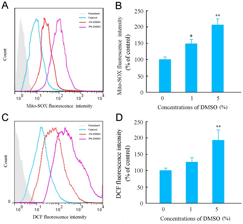
Figure 5. Effects of DMSO on mitochondrial and intracellular ROS generation in cultured mouse cortical astrocytes.
(A) Representative flow cytometry data showing Mito-SOX fluorescence, a highly selective indicator of superoxide in live cell mitochondria, in astrocytes treated with various concentrations of DMSO for 24 h. (B) Quantitative analysis revealed that the Mito-SOX fluorescence intensity increased in astrocytes treated with DMSO with a dose-dependent manner. (C) Representative flow cytometry data showing DCF fluorescence in astrocytes treated with various concentrations of DMSO for 24 h. (D) The quantitative analysis showed that ROS levels were increased in astrocytes treated with DMSO at 5% concentration but not at 1%, by detecting the fluorescence intensity of DCF. (E) Data are shown as a mean ± SEM of five independent experiments performed in triplicate. *P<0.05, **P<0.01 versus control group. #P<0.05, ## P<0.01 versus 5% DMSO treated group.

