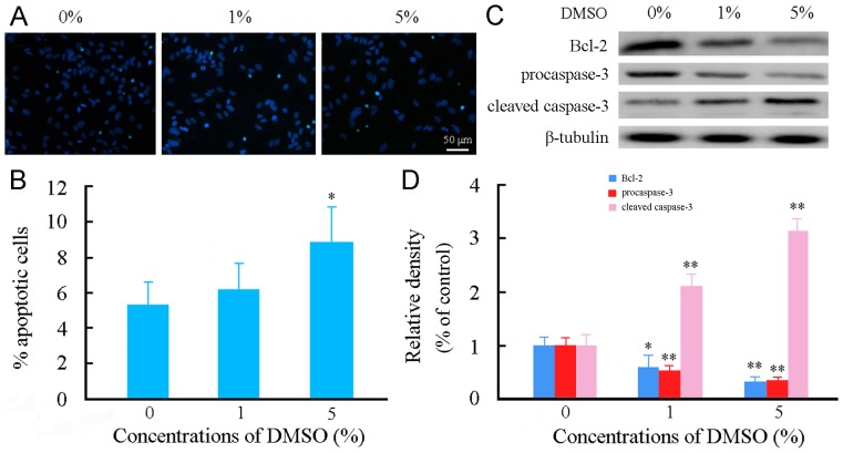
Figure 6. Effects of DMSO on apoptosis of cultured mouse cortical astrocytes.
Astrocytes were incubated with various concentrations of DMSO for 24 h. (A) Representative micrographs showing TUNEL positive apoptotic astrocytes (cyan-blue). Cell nuclei counterstained with Hoechst 33342 (blue). (B) Quantitative analysis of astrocyte apoptosis. (C) Representative western blot bands showing expression levels of procaspase-3, cleaved caspase-3 and Bcl-2 in astrocytes. (D) The quantitative analysis showed that Bcl-2 and procaspase-3 expression levels were decreased, but cleaved caspase-3 expression level was increased in astrocytes treated with 1% or 5% DMSO. Data are shown as a mean ± SEM of five (for TUNEL) or four (for Western blot) independent experiments performed in triplicate. *P<0.05 and **P<0.01 versus control group.

