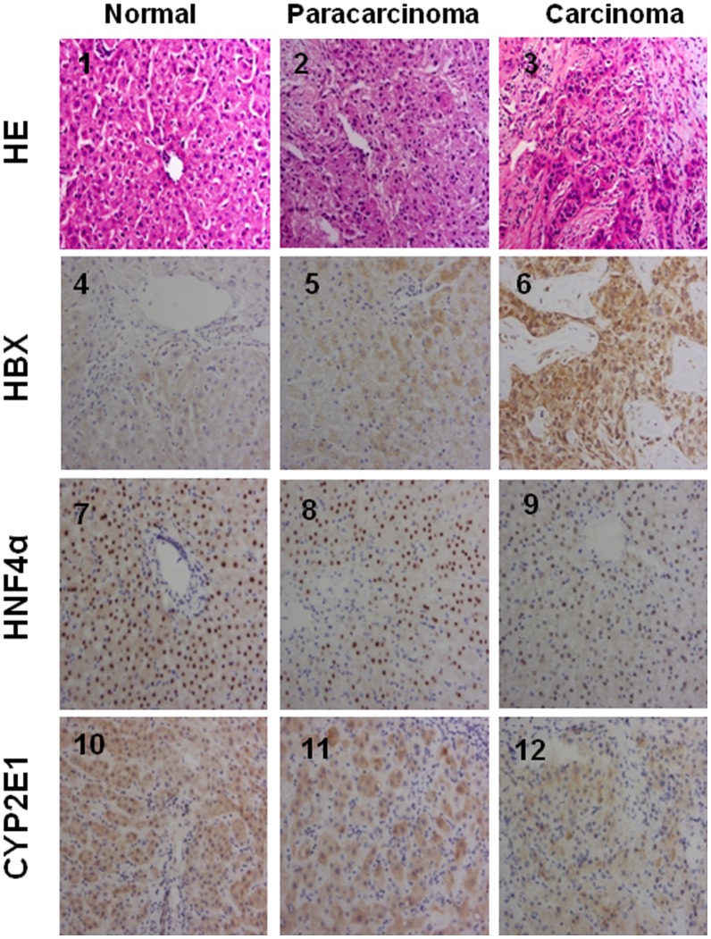
Figure 5. Immunohistochemical staining of HNF4α, CYP2E1 and HBx in human liver tissues.
Row 1, morphology observation with HE staining. Row 2, HBx immunochemical staining. Row 3, HNF4α immunochemical staining. Row 4, CYP2E1 immunochemical staining. All HE staining or immunochemical staining were shown as typical fields from five cases of human normal, and five cases of paracarcinoma or HCC liver tissues.

