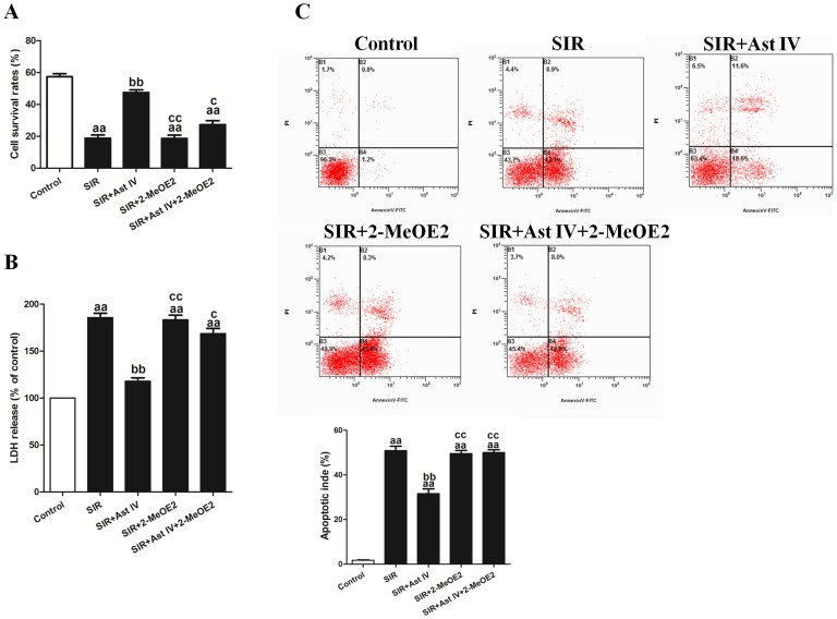
Figure 3. The effects of Ast IV and 2-MeOE2 post-ischemia treatment on cell viability, LDH release, and apoptotic index of SIR-injured cardiomyocytes.
(A). Cardiomyocyte viability was assessed using the MTT assay. (B). An ELISA assay was performed to detect the release of LDH in culture medium. (C). Representative flow cytometry apoptotic results are shown. Four subpopulations and their fractions are indicated: normal cells (lower left), dead cells (upper left), early apoptotic cells (lower right), and late apoptotic cells (upper right). The apoptotic index is expressed as the number of apoptotic cells/the total number of counted cells ×100%. The results are expressed as the mean±SEM, n = 6. aaP<0.01 vs. Control; bbP<0.01 vs. SIR; ccP<0.01 vs. SIR+Ast IV. SIR, simulated ischemia reperfusion; Ast IV, Astragaloside IV; 2-MeOE2, 2-methoxyestradiol; LDH, lactate dehydrogenase.

