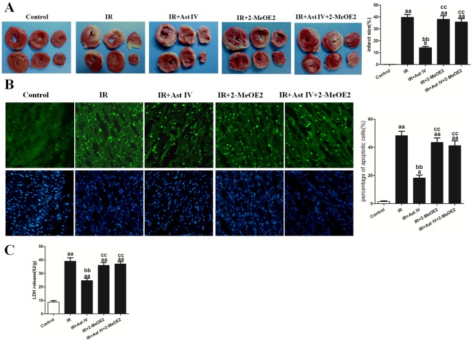
Figure 6. The effects of Ast IV and 2-MeOE2 post-ischemia treatment on the infarct size, apoptotic index, and LDH release of IR-injured isolated hearts.
(A). Representative images of the myocardial infarct size are shown. The infarction size is expressed as the percentage of infarct relative to the mass at risk. (B). Representative images of apoptotic cardiomyocytes are shown. The apoptotic cells were detected by immunofluorescent staining with TUNEL (green)and DAPI (blue) staining was used to label the nuclei. (C). The amount of LDH was normalized against the dry weight of the heart and is expressed as IU/g. The results are expressed as the mean±SEM, n = 6. aaP<0.01 vs. Control; bbP<0.01 vs. IR; ccP<0.01 vs. IR+Ast IV. IR, ischemia reperfusion; Ast IV, Astragaloside IV; 2-MeOE2, 2-methoxyestradiol; LDH, lactate dehydrogenase.

