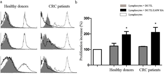
Figure 1. LMW HA treatment of DC enhances human T-cell proliferation in vivo.

Nude mice received intraperitoneal injections of human-derived CFSE-labeled PBLs (5×10⁶) and allogeneic mature DC/TL, or DC/TL/LMW HA (1,5×10⁶). Proliferation was monitored 48 h later by FACS-gated lymphocytes from peritoneal lavages by quantifying fluorescent dye signal which is inversely correlated to their proliferation rate. Fluorescence intensity in the input undivided lymphocytes was over 95% (gray line in the histogram). a) Three histograms representative of both HD and CRC patients are shown. Gray line: undivided lymphocytes; gray shadow: Lymphocytes + DC/TL; black line: Lymphocytes + DC/TL/LMW HA. b) Bars represent percentage of lymphocyte proliferation increase ± SEM respect to undivided lymphocytes (HD n: 5 and CRC patients n: 5). White bar: lymphocyte alone; gray bar: DC/TL; black bar: DC/TL/LMW HA. *DC/TL vs DC/TL/LMW HA; Paired t test; p≤0.05.
