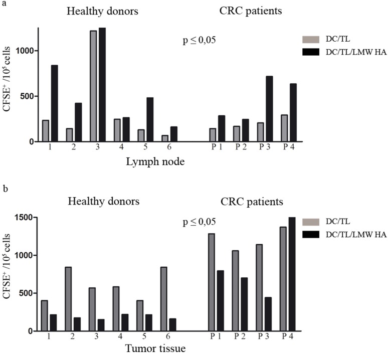
Figure 6. Pre-incubation of DC with LMW HA increases their migration toward lymph node and reduces their tumor attraction in vivo.
DC/TL were or not LMW HA treated, and after CM DiL labeling were inoculated s.c. in human-tumor bearing nude mice. Twenty four hours later lymph nodes (a) and tumors (b) were surgically removed and a cell suspension was obtained from these tissues. Number of fluorescent DC was counted by flow cytometry. The upper panel (a) shows DC/TL migration toward lymph nodes. The lower panel shows migration toward tumor tissue (b). Bars represent the number of migrated DC per individual HD (1–6) and CRC patients (P 1–4). Gray bar: DC/TL; black bar: DC/TL/LMW HA. * DC/TL vs DC/TL/LMW HA. Paired t test; *p≤0,05.

