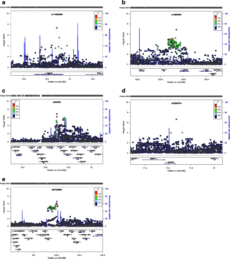
Figure 2.

The meta-analysis for total lung capacity measured by chest CT in COPD subjects. Regional association plots for (a) one genome-wide significant locus and (b-e) the other four suggestive loci. The x-axis is chromosomal position, and the y-axis shows the -log10 P value. The most significant SNP at each locus is labeled in purple, with other SNPs colored by degress of linkage disequilibrium (r2). Plots were generated using LocusZoom.
