
Fig. 2.
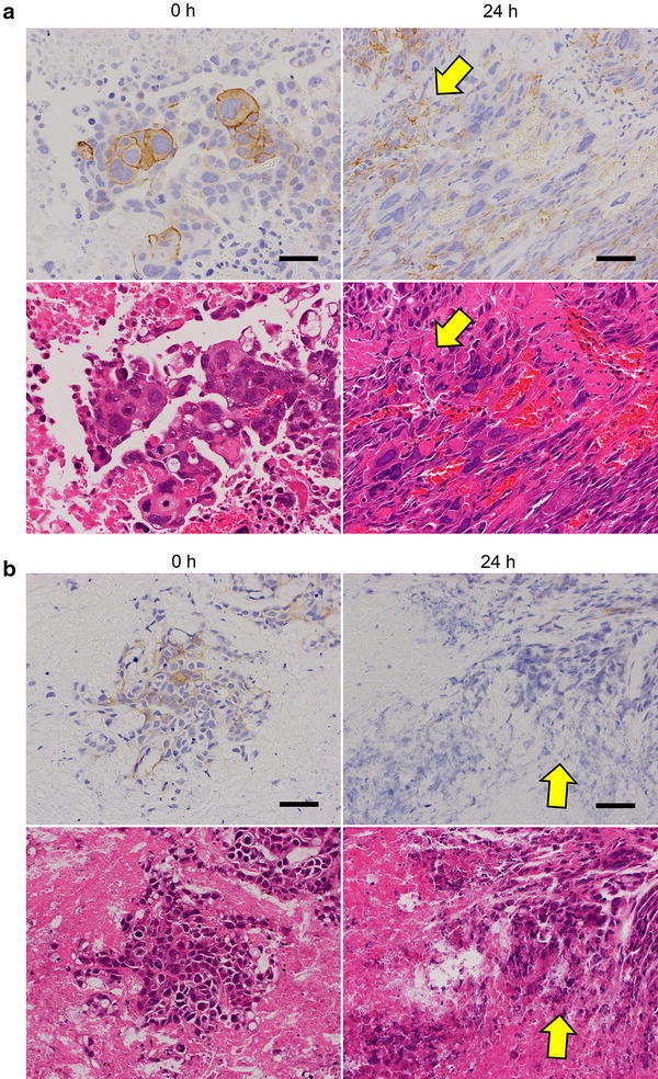
Effect of time to fixation on IHC staining for HER2 in tumor tissue center. Tumor tissue specimens of SCH (a) and (b) SNU-16 were collected and allowed to stand for 0 or 24 h before fixing with 10 % NBF for 24 h. Upper panels HER2 IHC staining; lower panels hematoxylin and eosin staining. Arrows indicate areas of advanced autolysis. Bars 50 μm