
Fig. 1.
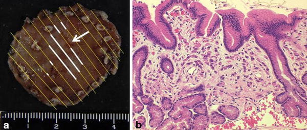
a Lesion resected with ESD. Resected specimen was 40 × 38 mm. The lesion was step-sectioned at 3- to 4-mm intervals, and then examined pathologically. Although SRCCs at the biopsy site disappeared (as an arrow indicates), the entire specimen revealed a lesion 13 × 13 mm in size (as white lines indicate). The resection was curative and the horizontal margin was 6.5 mm. b The specimen showed localized neoplastic cells with signet-ring features limited to the mucosal layer. Signet-ring cells are easily detected on Haematoxylin and eosin (H&E) sections