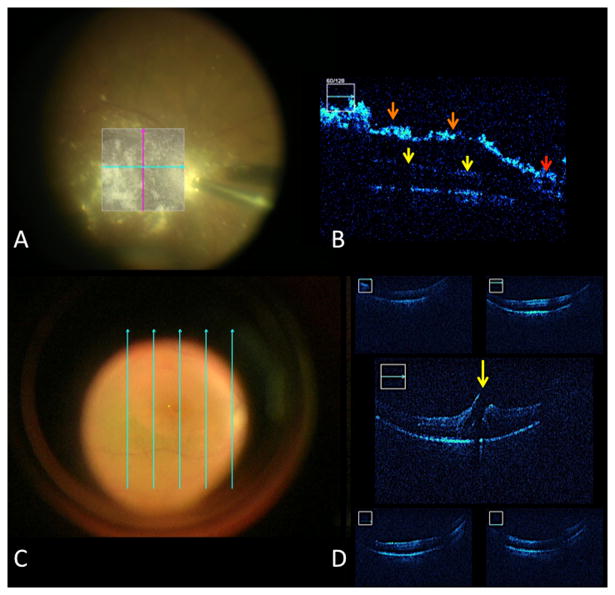
Figure 3.
Posterior segment intraoperative OCT (iOCT). (A) Surgical view following triamcinolone installation to visualize the hyaloid. (B) B-scan following OCT contrast enhancement with triamcinolone with excellent visualization of the posterior hyaloid (orange arrow) and attachment at the optic nerve (red arrow). Underlying retina is shadowed due to density of triamcinolone (yellow arrow). (C) Surgical view following internal limiting membrane (ILM) peeling with 5-line raster display. (D) Following ILM peeling, iOCT reveals persistence of the retinal flap and full-thickness macular hole (yellow arrow).

