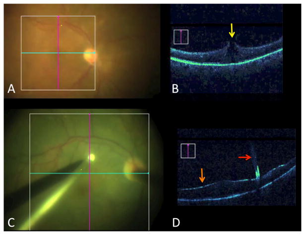
Figure 4.
Visualizing the impact of surgical maneuvers. (A) Surgical view with crosshairs of OCT scanner following elevation of the posterior hyaloid in a vitreomacular traction (VMT) case. (B) B-scan following hyaloid elevation in VMT case reveals occult full-thickness macular hole (yellow arrow), altering surgical planning (e.g., internal limiting membrane peeling, gas tamponade). (C) Surgical view of diamond dusted membrane scraper initiating membrane peel. (D) Real-time visualization with intraoperative OCT of instrument tissue-interaction (red arrow). Indocyanine green staining results in shadowing of underlying tissue and enhanced visualization of the internal limiting membrane (orange arrow).

