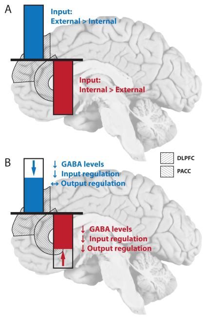
Figure 4. Resting state activity in PACC and DLPFC in controls and major depression.
(A) Imaging findings in healthy subjects show a reciprocal pattern of neural activity between PACC and DLPFC. Increased activity in DLPFC is accompanied by decreased activity levels in PACC and vice versa. The relative ratio of information content (Internal versus external) is depicted for both areas. (B) In MDD, the resting state activity of the DLPFC is reduced. In contrast the PACC shows increased resting state activity in MDD (i.e. less negative activity), resulting in an abnormal reciprocal modulation between the two regions. Underlying cellular and biochemical changes are highlighted and described in Figures 2-3.

