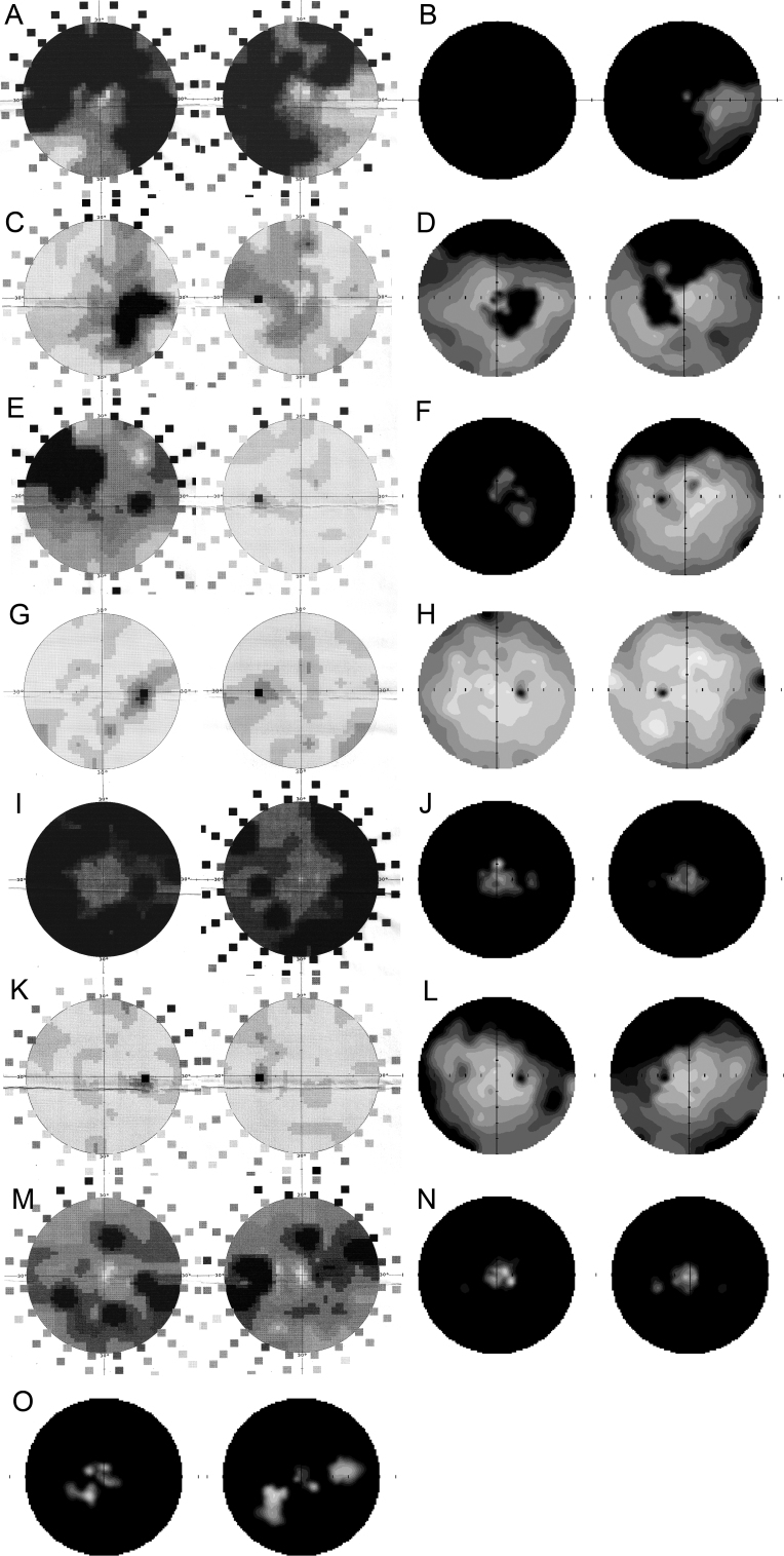
Figure 4.
Serial observations of disease progression on automated visual field for individuals with a c.2543del RPGR ORF15 mutation. Visual field scans (showing 50° nasal and temporal to fixation) of the right and left eyes of female III:4 at the age of 47 (A) and 58 (B) years; the right and left eyes of female III:1 at the age of 58 (C) and 64 (D) years; the right and left eyes of female III:9 at the age of 44 (E) and 50 (F) years; the right and left eyes of female IV:4 at the age of 25 (G) and 29 (H) years; the right and left eyes of male IV:3 at the age of 23 (I) and 30 (J) years; the right and left eyes of male IV:6 at the age of 14 (K) and 19 (L) years; the right and left eyes of male IV:2 at the age of 32 (M) and 38 (N) years; the right and left eyes of male IV:1 at the age of 42 (O) years. Static perimetry results are shown as 12-level gray scales of sensitivity loss. Black areas indicate no detection of stimuli. Physiologic blind spot is shown as a black circle at 12° in the temporal field.

