Figure 3.

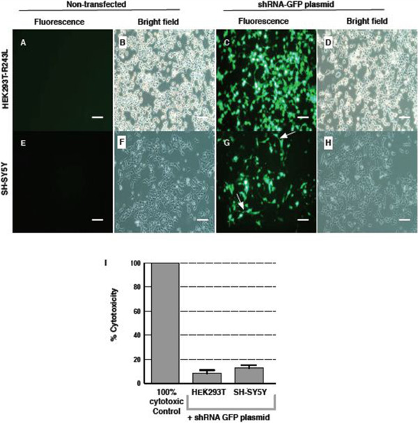
Cells express GFP after transfection with plasmids. We tested the transfection of shRNA-GFP plasmid in two cell types, HEK293T-CAPN5-p.R243L (A- D) and SH-SY5Y (E- H). C, G. HEK293T-CAPN5-p.R243L and SH-SY5Y neuroblastoma cells successfully transfected with negative control shRNA plasmid expressed GFP marker. In G. arrows show short neuritis extend from cell. A, E. Cells not transfected with plasmid show no background labeling under fluorescent light. B, D. Under bright field, HEK293T-CAPN5-p.R243L cells proliferate as an adherent layer. F, H. SH-SY5Y cells proliferate as an adherent clump. Cells were visualized under 20X magnification. Scale bars: 100 μm. I. As revealed by LDH assay, the knockdown of CAPN5 was not cytotoxic.
